Wisdom teeth (third molars) usually come through in the late teens and early twenties. When there is not enough room in the jaw, they cannot erupt fully and may remain partially or completely impacted.
This page explains when removal is considered, how the procedure is done and what the healing process looks like, for information purposes. Whether your tooth should be removed can only be determined through an examination and X-ray assessment.
Does every wisdom tooth need to be removed?
No. For impacted teeth that cause no symptoms and have no disease around them, monitoring is also an option. The decision takes into account:
- The position and angle of the tooth
- Whether there is recurrent inflammation around it
- Whether the tooth or the neighbouring tooth is decayed
- Whether a cyst or other pathology is visible on the X-ray
- Whether the area can be kept clean
- Any orthodontic treatment plan
- Age and general health
When is removal considered?
| Situation | Reason |
|---|---|
| Pericoronitis | Recurrent inflammation of the gum over a partially erupted tooth |
| Decay | Decay in the tooth itself or in the neighbouring molar that cannot be cleaned |
| Root resorption | The impacted tooth causing resorption of the neighbouring tooth's root |
| Cyst / pathology | A sac-like lesion around the tooth |
| Gum pocket | A deep pocket behind the neighbouring tooth that cannot be cleaned |
| Orthodontics | Situations required by the treatment plan |
How is the removal carried out?
- Examination and imaging. The position of the tooth, the shape of its roots and its distance to the nerve canal in the lower jaw or the sinus in the upper jaw are assessed with a panoramic X-ray and, where necessary, a CT scan.
- Planning. It is decided whether a simple or a surgical extraction is needed; medication and general health are reviewed.
- Anaesthesia. The area is numbed with local anaesthesia.
- Access. For impacted teeth a flap is opened in the gum; if necessary, some of the bone covering the tooth is removed.
- Removal. The tooth is removed in one piece or in sections.
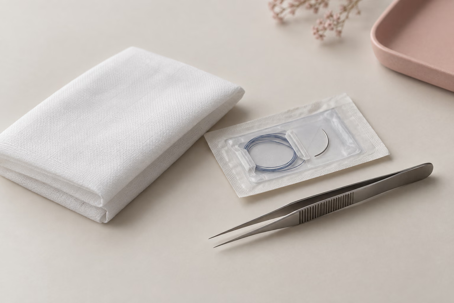
- Cleaning and closure. The area is rinsed and stitches are placed if needed.
- Follow-up. Stitches are usually removed after about one week.
The healing process
| Period | What to expect |
|---|---|
| First 24 hours | Oozing; protecting the blood clot is important |
| Days 2–3 | Swelling is at its most noticeable |
| Days 4–7 | Swelling and pain decrease markedly |
| Around day 7 | Stitches are removed |
| 2–3 weeks | The gum has largely closed |
| A few months | The bone cavity fills in |
Aftercare
What to avoid in the first 24 hours:
- Spitting or rinsing vigorously
- Drinking through a straw
- Smoking
- Hot food and drinks
- Touching the area with your tongue or fingers
What is recommended:
- Biting on the gauze placed by the dentist for the time advised
- Applying cold to the cheek from outside, at intervals
- Eating soft, lukewarm foods
- Taking prescribed medication exactly as instructed
- From the next day, starting gentle cleaning as your dentist instructs
Risks and complications
- Infection at the surgical site
- Dry socket: loss of the blood clot and severe pain
- Prolonged or recurrent bleeding
- Swelling, bruising and temporarily limited mouth opening
- Damage to the neighbouring tooth or its filling
- Numbness of the lip/chin area due to the proximity of the nerve in the lower jaw — usually temporary, rarely permanent
- Problems related to the sinus in the upper jaw
- Jaw fracture (very rare)
This list is not exhaustive. Risks specific to you are discussed in detail during the informed consent process, based on the position of the tooth on the X-ray.
When should I contact my dentist?
- Pain that increases after the third day and is not relieved by medication
- Bleeding that does not stop
- High fever and widespread swelling
- Increasing difficulty opening your mouth
- A bad smell and bad taste in the mouth
In an emergency, call 112.
What should patients travelling from abroad consider?
The removal itself takes one visit, but swelling peaks on days 2–3 and stitches are usually removed after about seven days. Whether the tooth should be removed at all is decided only after an examination and imaging (panoramic X-ray and, where necessary, a CT scan).
| Topic | What to know |
|---|---|
| Procedure | About 20–60 minutes, depending on the position of the tooth |
| First days | Swelling is most noticeable on days 2–3; returning to work usually takes 1 day after a simple and 2–3 days after a surgical extraction |
| Stitches | Usually removed after about 7 days |
| Records in advance | Existing panoramic X-rays or CT scans can be shared before your visit |
| Back home | Stitch removal and any later check-ups can be arranged with a local dentist |
About treatment fees
No fee information is given on this page. The scope of the procedure depends on the position of the tooth and how deeply it is impacted; fees can only be discussed after an examination and X-ray assessment.